
Arterial Ultrasound of Legs: What to Expect From Your PAD Tests
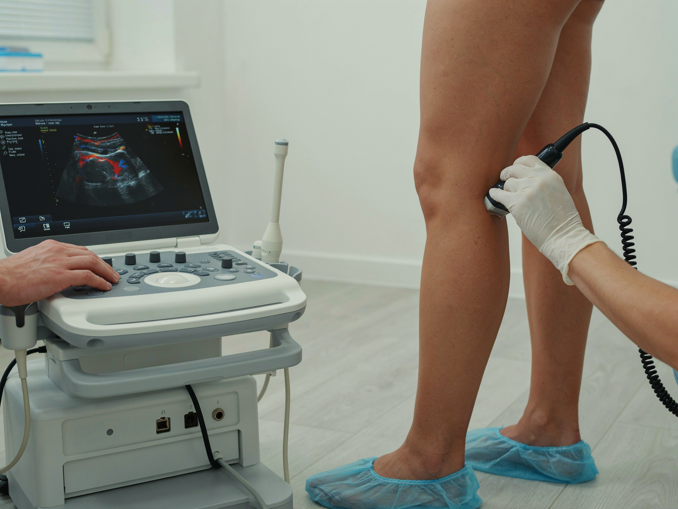
Your doctor says you need an arterial ultrasound of legs, and the questions start right away. Will it hurt? How long will it take? What happens if they find a blockage? Feeling nervous before a medical test is normal, especially when you don't know what to expect.
So let's start with some reassurance. An arterial ultrasound is one of the gentlest tests in medicine. There are no needles, no dye, and no radiation. It's warm gel, a small handheld probe, and a detailed look at how blood moves through your legs.
In this guide, we explain what happens at your first consultation, how each test for peripheral artery disease (PAD) works, what your results mean, and how to prepare so you leave with clear answers.
What to Expect During Your Initial Consultation
Every good diagnosis starts with a conversation. Before any testing, we'll sit down with you and ask questions like these:
- Where do you feel pain or discomfort, and what does it feel like?
- How far can you walk before symptoms start, and how long do they take to ease?
- Do your feet feel cold or numb? Do you have sores that are slow to heal?
- Do you smoke or vape now, or have you in the past?
- Do you have diabetes, high blood pressure, high cholesterol, or kidney disease?
- Has anyone in your family had PAD, a heart attack, or a stroke?
Next comes a focused physical exam. We check the pulses in your groin, behind your knees, on the tops of your feet, and behind your ankle bones. We look at your skin color, temperature, hair growth, and nails, and we check for any wounds. We may listen over your arteries with a stethoscope for a bruit, a whooshing sound that can signal turbulent flow through a narrowed artery. We may also raise your legs for a minute to see whether your feet turn pale, then lower them to see how quickly the color returns.
This first step tells us which tests you actually need, so you don't go through anything unnecessary.
Common Diagnostic Tests Explained
1. Non-Invasive Imaging and Scans
Arterial duplex ultrasound. This is the test most people mean when they say "arterial ultrasound of legs." You may also see it called a lower extremity arterial Doppler. It combines two kinds of ultrasound in one exam:
- B-mode imaging shows a picture of the artery wall and any plaque inside it.
- Doppler measures how fast, and in which direction, blood is flowing.
When blood squeezes through a narrowed spot, it speeds up, like water through a pinched garden hose. If the speed at one spot is roughly double the speed just above it, that usually means the artery is narrowed by 50% or more. The Doppler also records the shape of each pulse. A healthy artery produces a crisp, multi-phase waveform. Past a blockage, the waveform turns flat and sluggish.
How it feels: You'll lie on an exam table in a gown or loose shorts. A sonographer spreads warm gel on your skin and glides a small probe from your groin down to your ankle. You may be asked to turn on your side or stomach so we can see behind the knee. The probe presses firmly at times, especially at the groin, but it shouldn't hurt. A study of both legs usually takes about 30 to 60 minutes. There's no recovery time, and you can drive yourself home.
CT angiography (CTA runoff) and MR angiography (MRA). If a procedure is being considered, we may need a more detailed road map from your abdomen to your toes. A CTA runoff uses a CT scanner and contrast dye given through an IV. The scan itself takes only minutes, and you may feel a brief warm flush when the dye goes in. Because the dye passes through your kidneys, we check your kidney function first. An MRA uses a magnetic field instead of X-rays, along with a different type of contrast. It takes longer and isn't right for everyone, including some people with certain implants or advanced kidney disease.
2. Specialized Diagnostic Testing
Exercise ABI (treadmill test). Some people have classic leg pain but a normal resting test. A partial blockage may let enough blood through at rest, but not during activity. With an exercise ABI test, we measure your ankle and arm pressures at rest, have you walk on a treadmill at a set pace and incline until symptoms stop you or you reach a time limit, then measure again. A significant drop in ankle pressure after exercise confirms PAD.
Pulse volume recording (PVR test) with segmental pressures. For a PVR test, we place blood pressure cuffs at several levels on each leg, typically the thigh, calf, and ankle. Each cuff is inflated slightly to record the small change in leg volume with every heartbeat. The shape of those waveforms, and the pressure drop between levels, helps pinpoint where a blockage sits. PVR is especially useful when arteries are stiff or calcified.
Toe-brachial index (TBI). In people with diabetes or kidney disease, the arteries at the ankle can become so stiff that a cuff can't compress them, which produces a falsely high ABI (above 1.40). Toe arteries are usually less affected, so we use a small cuff on the big toe instead. A TBI of 0.70 or lower points to PAD.
Blood work. Lab tests don't diagnose PAD directly, but they help explain why it developed and guide treatment. Common tests include a lipid panel, A1C for blood sugar, and kidney function.
3. In-Office Clinical Assessments
Resting ankle-brachial index (ABI). This is usually the first test we do, and the whole thing typically takes 15 to 20 minutes, including rest time. After you lie on your back for several minutes, we place blood pressure cuffs on both arms and both ankles and use a small handheld Doppler to listen for the pulse. Your ABI is your ankle pressure divided by your arm pressure. Results are read like this:
- 1.00 to 1.40: Normal
- 0.91 to 0.99: Borderline
- 0.90 or lower: PAD is present. Lower numbers mean more severe disease.
- Above 1.40: Stiff, calcified arteries. We'll usually add a toe pressure test.
Other quick bedside checks include a pulse exam, capillary refill (how fast color returns after gently pressing on a toenail), and a careful inspection of your feet.
How Diagnostic Results Guide Your Treatment Plan
Test results take the guesswork out of your care. They typically shape your next steps like this:
- Normal results: If your arteries look healthy, we look for other explanations, such as spinal stenosis, nerve problems, arthritis, or vein disease.
- Mild to moderate PAD: Most people start with a structured walking program, a statin, an antiplatelet medicine, blood pressure and blood sugar control, and help quitting tobacco. We repeat testing over time to track your progress.
- PAD that limits your life or threatens your leg: If walking pain doesn't improve with treatment, or if you have rest pain or wounds, we use your ultrasound or CTA to plan a procedure that restores blood flow.
When a Peripheral Angiogram Is Needed
A peripheral angiogram gives the most detailed picture of your leg arteries, and it's usually done when treatment is being planned. During a leg angiogram procedure, you receive local numbing medicine and light sedation. A thin catheter is guided through a small puncture, usually in the groin or wrist, and contrast dye is injected while X-ray images are taken. It often takes about an hour. One big advantage: if we find a blockage, we can often treat it in the same session with angioplasty or a stent. Many patients go home the same day after a few hours of rest.
Your results map directly to your options. You can explore the full range of PAD treatments at Healthy Living Heart and Vein, from medication and walking programs to angioplasty, stenting, bypass surgery, and endarterectomy.
How to Prepare for Your Diagnostic Visit
A little preparation makes your visit smoother and your results more accurate:
- Dress for easy access. Wear loose, comfortable clothes. Shorts or pants that roll up easily are ideal.
- Ask about fasting. Most leg artery ultrasounds need no special prep. If your abdominal arteries are included, you may be asked not to eat for several hours beforehand to reduce bowel gas. If you have diabetes, ask how to handle your medicines.
- Skip tobacco and caffeine beforehand. Both can temporarily affect your blood vessels and blood pressure readings. Follow your testing center's instructions.
- Bring your records. Bring a list of your medicines, any past vascular test results, and details about prior procedures or stents.
- Tell us about allergies and kidney issues. This matters if contrast dye may be used for a CTA or angiogram.
- Track your symptoms. Note where you feel pain, how far you can walk before it starts, and how long it takes to ease.
Questions to ask at your visit:
- What is my ABI, and what does it mean?
- Where are my blockages, and how severe are they?
- Do I need more testing, or do we have enough information?
- What are my treatment options, and what do you recommend first?
- How often should my arteries be rechecked?
Get Clear Answers About Your Health
Not knowing what's causing your leg pain is often harder than the diagnosis itself. The right tests replace worry with a plan. An arterial ultrasound of legs is safe, painless, and one of the best tools we have to see exactly what's happening inside your arteries.

Book Your Comprehensive Diagnostic Evaluation
Healthy Living Heart and Vein offers same-week appointments for patients in Conroe, The Woodlands, Huntsville, Willowbrook, and Spring, Texas. Call (936) 978-0466 to get started.
Book Your EvaluationSources
- Collins R, et al. Duplex Ultrasonography, MRA, and CTA for Diagnosis and Assessment of Symptomatic Lower Limb PAD: Systematic Review. BMJ, 2007
- 2024 ACC/AHA Guideline for the Management of Lower Extremity Peripheral Artery Disease (Circulation)
- CDC: About Peripheral Arterial Disease (PAD)
Medical Disclaimer: This content is for educational purposes only and does not constitute formal medical advice, diagnosis, or treatment. Always follow the preparation instructions given by your own care team.